Development Sequence
Early Chick Development, 24 Hr. Chick, and 72 Hr. Chick
Neural_groove.jpg
Neural Groove

24 Hr. Transverse labels.jpg
24 Hr. Transverse labels

72 HR. 10X.jpg
72 HR. 10X

72 Hr. 40X.jpg
72 Hr. 40X

72 HR. 4X.jpg
72 HR. 4X

Mesoderm derivatives.jpg
Mesoderm derivatives

Transverse.jpg
Transverse
1
of
7
-
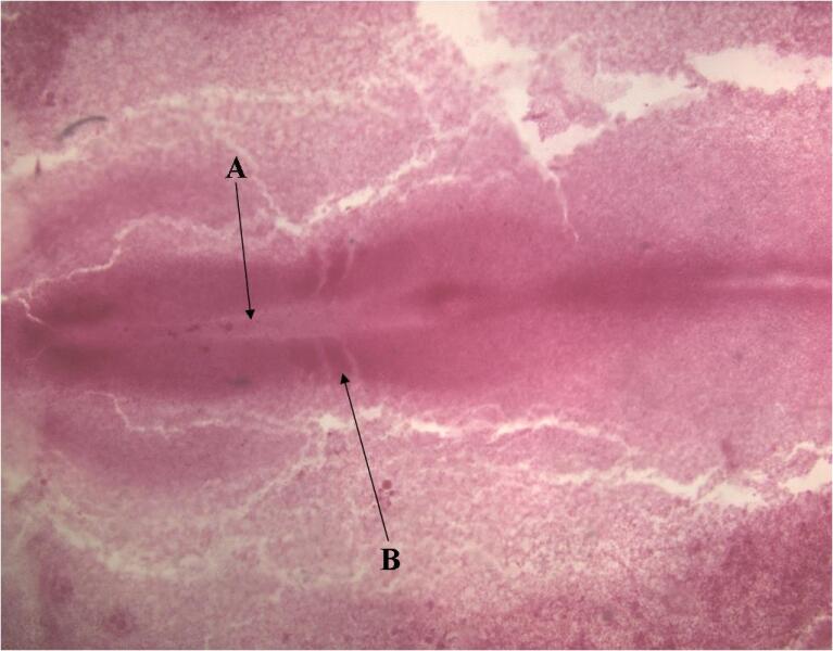
Toggle ItemThe Neural GrooveA: The Neural Groove is the precursor to the neural tube which will eventually give rise to the central nervous system. The neural groove forms when the ectoderm thickens, and starts to invaginate causing a large groove down the length of the embryo on the distal side.
B: The Somites are segmented blocks of mesoderm that form on either side of the neural tube they give rise to body and limb muscles, cartilage that forms the vertebrae and ribs, and the dermis. -
Toggle Item24 Hr. Transverse LabelsA: The ectoderm here is located dorsal to the whole embryo.
B: The lateral plate mesoderm is usually split into two separate layers: the splanchnic and somatic mesoderm
C: The endoderm is much thinner than the mesoderm or the ectoderm is also located just beneath the notochord
D: The neural tube is located at the dorsal axis and is derived from ectoderm it eventually becomes the central nervous system.
E: The notochord is located just beneath the neural tube and is in charge of providing support for the embryo. It is derived from the mesoderm called cohordamesoderm.
F: The dermamyotome is located on the dorsal edge of the somite
G: The scleratome is located just beneath the dermamyotome on the ventral edge of the somite
H: Somatic mesoderm is on the dorsal side of the lateral plate mesoderm
I: Splanchnic mesoderm is on the ventral side of the lateral plate mesoderm -
Toggle ItemTransverseA: The Ectoderm layer is characterized by how compact and closely together the cells are. The cells have little or no space in between. At this stage sometimes the ectoderm has started to thicken and a groove forms in its center called the neural groove which later becomes the neural tube
B: The Mesoderm layer is about the same thickness but you will notice it has more loosely packed or mesenchymal cells. The mesoderm is also easily identified because it is located between the two other layers. Mesoderm gives rise to much of the musculoskeletal system.
C: The Endoderm layer is almost always the thinnest layer of the three and it gives rise to much of the internal organs of the body especially the digestive tract. -
Toggle ItemMesoderm DerivativesA: Somatic Mesoderm
B: Dermamyotome
C: The intermediate mesoderm is located between the paraxial mesoderm (dermamyotome and scleratome) and the lateral plate mesoderm. The intermediate mesoderm gives rise to the kidney and the gonads. This can be easily remembered because if these two intermediate mesoderm tissues drop straight down the body they would end up where the kidneys and gonads would be.
D: Scleratome. You can see the slight differentiation between the dermamyotome and the scleratome.
E: Splanchnic mesoderm -
Toggle Item72 Hr. 10XA: Neural Tube
B: The Notochord tissue looks just like the rest of the mesoderm, but the tissue inside the dorsal aorta has many red blood cells. This is the easiest way to differentiate the two
C: The dorsal aorta has red blood cells inside its lumen
D: The gut endoderm is the thick ring of cells on the interior. This will eventually become the epithelial layer lining the GI tract -
Toggle Item72 Hr. 40XA: Neural tube
B: Neural Coele
C: Notochord
D: Dorsal Aorta
E: Gut endoderm -
Toggle Item72 Hr. 4XA: The neural tube is completely closed and the brain is beginning to form the neural tube cells are tightly compacted.
B: The notochord gives stability to the embryo as it grows and then it degenerates.
C: The dorsal aorta is located just beneath the notochord and is usually seen as one or two large cavities filled with red blood cells. This will later become the mature heart
D: The gut endoderm can be recognized by the thick compact ring of epithelium located within a lumen below the dorsal aorta. The lumen is also usually surrounded by a large amount of other tissue